
How Mammography Works: Types, Process, and Importance
Introduction to Mammography
Mammography is a specialized medical imaging technique that uses low-dose X-rays to visualize the internal structures of the breast. It is considered the gold standard for breast cancer screening and detection. By producing detailed images of the breast tissue, mammography allows healthcare professionals to identify potential abnormalities, such as tumors or cysts, that may indicate the presence of breast cancer.
This condition is one of the most prevalent types of cancer among women, and early detection is critical for successful treatment, that is why Mammography plays a vital role, as it can identify abnormalities even before they are palpable or cause noticeable symptoms. Regular screening mammograms can detect breast cancer at an early stage when it is more likely to be curable.
The American Cancer Society and the National Cancer Institute recommend that women undergo regular screening mammograms starting at the age of 40. However, the frequency and timing of mammograms may vary depending on individual risk factors and guidelines from healthcare providers. Mammography is not only used for screening but also for diagnostic purposes when a woman presents with a breast problem or an abnormality is detected during a screening mammogram.
In recent years, advancements in mammography technology have further improved its diagnostic capabilities. Digital mammography, for example, allows for the acquisition of high-resolution digital images, which can be stored, manipulated, and transmitted electronically.
Mammography is typically performed in dedicated mammography facilities or breast imaging centers, which have specialized equipment and highly trained personnel. These facilities adhere to strict quality standards to ensure accurate and reliable results.
The Mammography Quality Standards Act (MQSA) in the United States, for instance, sets guidelines for mammography units, technologists, and interpreting physicians, aiming to maintain high standards of quality and patient care. Mammography is typically performed in dedicated mammography facilities or breast imaging centers, which have specialized equipment and highly trained personnel. These facilities adhere to strict quality standards to ensure accurate and reliable results.

Mammography is typically performed in dedicated mammography facilities or breast imaging centers, which have specialized equipment and highly trained personnel. These facilities adhere to strict quality standards to ensure accurate and reliable results. The Mammography Quality Standards Act (MQSA) in the United States, for instance, sets guidelines for mammography units, technologists, and interpreting physicians, aiming to maintain high standards of quality and patient care.
Types of Mammograms:
Mammography has evolved over the years, offering different types of imaging techniques to cater to varying needs and circumstances. Here are the primary types of mammograms commonly used in breast imaging:
- Screening Mammogram: A screening mammogram is a routine examination performed on women who do not exhibit any specific breast problems or symptoms. It is typically conducted on an annual basis or as recommended by healthcare providers. The screening mammogram aims to detect any early signs of breast cancer before symptoms become noticeable. It involves obtaining two-dimensional (2D) X-ray images of the breast from different angles, providing a comprehensive view of the breast tissue.
- Diagnostic Mammogram: A diagnostic mammogram is performed when further evaluation is required after an abnormality is detected during a screening mammogram or when a woman presents with specific breast concerns. This type of mammogram involves additional views and specialized imaging techniques to obtain more detailed images of the breast. Diagnostic mammograms are highly useful in evaluating suspicious areas, determining the nature of a detected abnormality, and guiding further investigations or interventions.
- 3D Mammography: Digital breast tomosynthesis, commonly referred to as 3D mammography, is an advanced imaging technique that provides a three-dimensional view of the breast. Unlike traditional 2D mammograms, Digital Breast Tomosynthesis captures multiple X-ray images of the breast from different angles, allowing for more precise evaluation of breast tissue. This technology has shown promising results in improving the detection of small tumors and reducing false positives, especially in women with dense breasts.
Procedure and Preparation:
Undergoing a mammogram requires minimal preparation and is generally a straightforward procedure. Here’s what you can expect:
- Scheduling the Mammogram: You can schedule a mammogram through a primary care physician or directly with a breast imaging center or mammography facility. It is advisable to inform the facility if you have breast implants or any specific concerns regarding breast health.
- Preparation: On the day of the mammogram, it is recommended to avoid using deodorants, perfumes, powders, or any products on the underarm or chest area. These substances can interfere with the imaging process. Additionally, it is preferable to wear a two-piece outfit to make undressing from the waist up more convenient during the procedure.
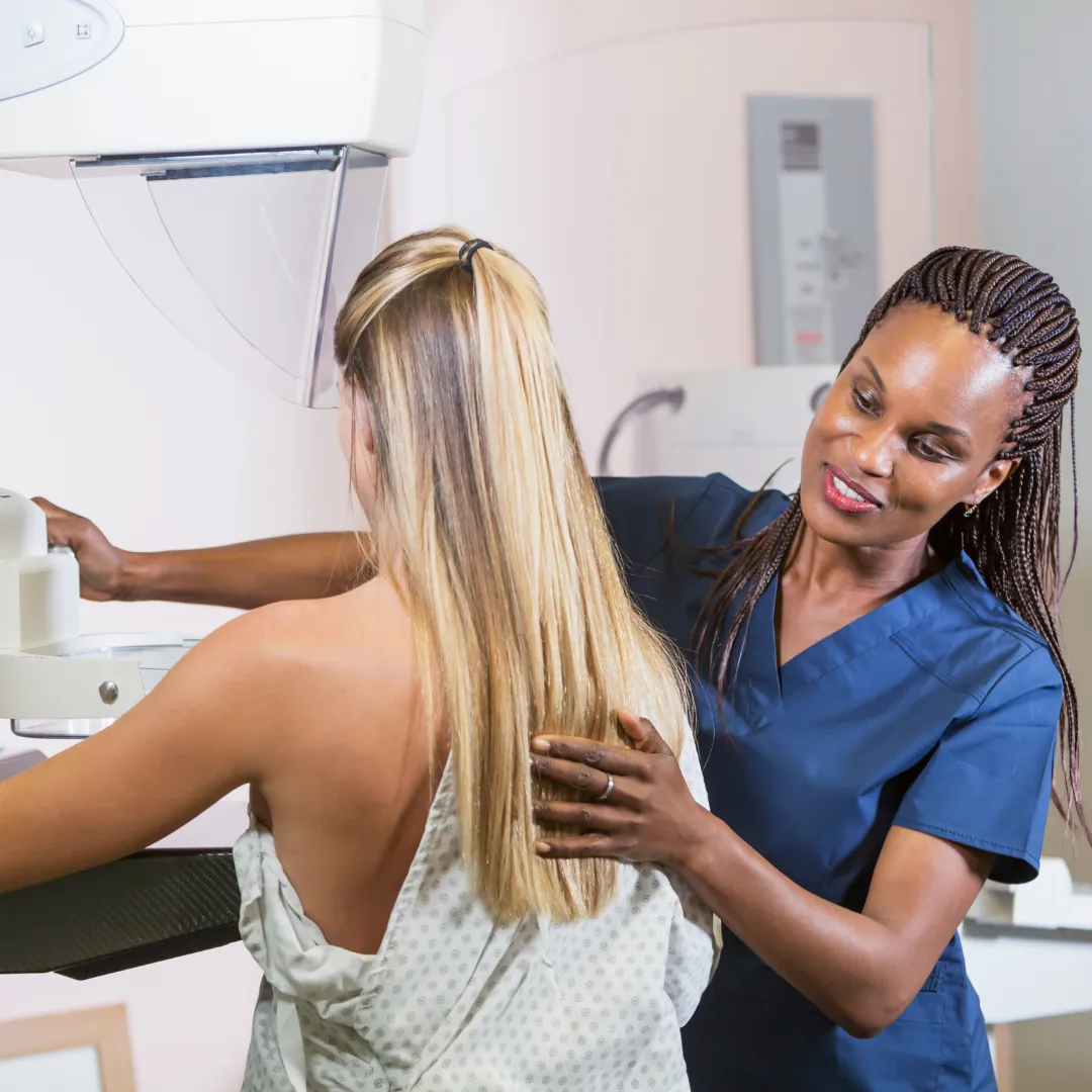
- The Mammography Procedure: During the mammogram, you will be positioned in front of a specialized mammography unit. A technologist, trained in performing mammograms, will assist you throughout the process. The breast tissue will be compressed between two plates to spread it out and obtain clear images. Compression is necessary to minimize motion and improve image quality.
- Image Acquisition and Interpretation: The technologist will position your breasts for imaging. They will then step behind a protective shield while an X-ray machine captures images of the breast from different angles. The process will be repeated for each breast. The acquired images will be interpreted by a radiologist, who specializes in diagnosing breast abnormalities, to identify any potential areas of concern.
- Follow-up and Results: Once the mammogram is complete, you will be provided with information on when and how you can expect to receive the results. If the mammogram raises any concerns, further diagnostic tests or evaluations may be recommended. Conversely, if the mammogram appears normal, you will be advised on when to schedule your next routine screening mammogram.

Importance of Mammography for Early Detection:
As you read before, mammography plays a vital role in the early detection of breast cancer, providing several significant benefits that contribute to improved outcomes and survival rates. Here are key reasons why mammography is essential for early detection:
- Detecting Abnormalities before Symptoms Arise: Breast cancer often presents no noticeable symptoms in its early stages. Mammography can detect small tumors or suspicious areas before they can be felt by hand or cause visible changes in the breast. By identifying these abnormalities early, healthcare providers can initiate prompt and targeted interventions, improving the chances of successful treatment.
- Increased Survival Rates: Early detection through mammography is associated with increased survival rates for breast cancer patients. When breast cancer is diagnosed at an early stage, the available treatment options are often more effective, and the likelihood of achieving a cure is higher. Regular mammograms enable the detection of cancer in its earliest stages, when it is most treatable.
- Screening for High-Risk Individuals: Mammography is particularly crucial for individuals at high risk of developing breast cancer. This includes women with a family history of the disease, certain genetic mutations, or other risk factors. For high-risk individuals, mammograms may begin at an earlier age or be conducted more frequently to maximize the chances of early detection and intervention.
- Tracking Changes over Time: Routine breast screenings provide a reference point for tracking changes in breast tissue over time. By comparing images from previous screenings, radiologists can identify any new developments, subtle changes, or abnormalities that may require further investigation. This longitudinal approach allows for a comprehensive evaluation of breast health and early detection of potential issues.
Addressing Concerns and Common Myths:
Despite the proven benefits of mammography, there are concerns and myths surrounding this breast imaging technique. Let’s address some of these concerns and provide clarity on common misconceptions:
- Discomfort and Compression: One concern often raised about mammograms is the discomfort associated with breast compression. While the compression may cause temporary discomfort, it is necessary to spread out the breast tissue and obtain clear images. The compression lasts only for a few seconds, and the potential benefits of early detection outweigh the temporary discomfort.
- Radiation Exposure: Mammography uses low-dose X-rays to capture images of the breast. The radiation exposure during a mammogram is minimal and considered safe. The benefits of early detection far outweigh any potential risks associated with the low levels of radiation involved in the procedure.
- False Positives and Overdiagnosis: Mammography can occasionally yield false-positive results, leading to further evaluations or biopsies. However, false-positive results are relatively uncommon, and additional diagnostic tests can help determine whether further intervention is necessary. Overdiagnosis, while a concern, is minimized through rigorous quality standards and the expertise of interpreting radiologists.
- Efficacy in Dense Breasts tissue: Women with this condition may be concerned about the accuracy of mammograms. Dense breast tissue can make it more challenging to detect abnormalities on a mammogram. However, supplemental screening options, such as breast ultrasound or breast MRI, can be employed to enhance the accuracy in the results.
- Alternative Screening Methods: While other imaging modalities, such as breast MRI or breast ultrasound, have their roles, mammography remains the primary screening tool for breast cancer. These alternative methods are often utilized as supplemental tests for further evaluation or in specific cases where mammography may not provide optimal results.
Recommendations and Frequency:
Breast cancer screening guidelines provide recommendations on when and how often women should undergo mammography. These recommendations may vary depending on factors such as age, personal medical history, and risk factors. Here are some general guidelines:
- American Cancer Society (ACS) Recommendations: The ACS suggests that women with an average risk of breast cancer begin annual mammograms at age 40. They emphasize the importance of yearly screenings, as opposed to less frequent intervals, to maximize the chances of early detection. However, they also acknowledge the importance of shared decision-making between women and their healthcare providers, considering individual preferences and values.
- U.S. Preventive Services Task Force (USPSTF) Recommendations: The USPSTF recommends biennial mammography screening for women aged 50 to 74. They suggest that women aged 40 to 49 make an informed decision with their healthcare providers about the potential benefits and risks of mammography. Additionally, they highlight the need for personalized screening decisions based on individual risk profiles.
- High-Risk Individuals: Women at high risk of developing breast cancer may require more frequent or earlier mammography screenings. This includes individuals with a strong family history of breast cancer, certain genetic mutations (such as BRCA1 or BRCA2), or previous chest radiation therapy.
It is important for women to discuss their specific risk factors and medical history with their healthcare provider, who can provide personalized recommendations regarding mammography frequency and starting age.
Regular screenings offer the best opportunity for detecting breast cancer at its earliest and most treatable stage. While concerns and misconceptions exist, addressing them through education and open communication is crucial to encourage women to undergo recommended screenings.
By adhering to guidelines and working closely with healthcare providers, women can make informed decisions about the health of their breast. Mammography, along with advancements in breast imaging technology, provides a powerful tool in the fight against breast cancer. Through regular screenings, we can increase the likelihood of early detection, improve treatment outcomes, and ultimately save lives.
Breast cancer awareness month serves as a reminder of the importance of mammography and the significance of early detection. By prioritizing regular mammograms and promoting the breast care, we can make a significant impact in reducing the burden of breast cancer and improving the well-being of women around the world.